

CBCT Panoramic Comparison for Dental Practices
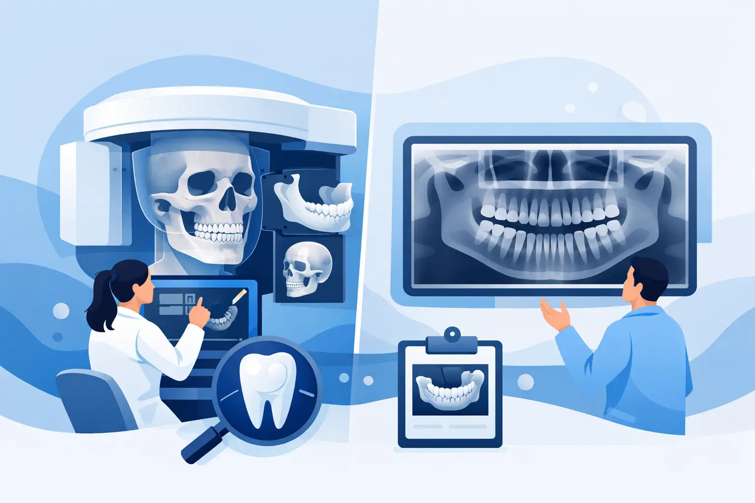
A CBCT panoramic comparison is not simply a question of choosing between a 2D image and a 3D image. It is a decision about diagnostic confidence, appointment flow, referral patterns, and how often your team needs information that a conventional panoramic study cannot provide. For a practice adding imaging capacity or replacing aging equipment, the best choice depends on the procedures you perform, the patients you serve, and the clinical questions you need to answer every week.
A panoramic unit remains an efficient, familiar workhorse. CBCT adds dimensional information that can change treatment planning in implants, endodontics, surgery, orthodontics, and complex restorative cases. Neither system is automatically the better purchase for every office. The practical value comes from matching imaging capability to real clinical demand.
CBCT Panoramic Comparison: 2D Versus 3D Information
Panoramic radiography captures a broad two-dimensional view of the maxillofacial region in one exposure. It is commonly used for new-patient exams, dentition overview, eruption assessment, third-molar screening, gross pathology review, and baseline documentation. A pano is fast to acquire, straightforward for staff to operate, and easy to incorporate into a routine hygiene or comprehensive exam workflow.
Its limitation is inherent to the format. Three-dimensional anatomy is compressed into a flat image. Structures may be magnified, distorted, superimposed, or obscured by positioning and motion. When a clinician needs to determine the buccolingual position of an impacted tooth, the relationship between an implant site and the inferior alveolar canal, or the true extent of a periapical lesion, a panoramic image may raise the question without resolving it.
CBCT uses a cone-shaped X-ray beam and reconstruction software to create a volumetric dataset. The clinician can review axial, coronal, sagittal, cross-sectional, and reconstructed panoramic views, subject to the system’s software and selected field of view. Rather than estimating where anatomy sits in space, the provider can evaluate it directly.
That extra information is valuable only when it affects care. CBCT should not replace panoramic imaging for every patient or become a default screening study. Good imaging protocols follow the ALARA principle - radiation exposure should be as low as reasonably achievable - while still providing the diagnostic information needed for the case.
When a Panoramic System Makes the Most Sense
For practices focused primarily on preventive, general, and straightforward restorative dentistry, a dedicated panoramic unit can deliver excellent everyday value. It supports efficient new-patient records, routine diagnostic assessment, third-molar monitoring, and broad treatment planning without adding the acquisition cost, file management demands, or training requirements of a 3D platform.
A panoramic system is also a sensible choice when a practice has reliable access to local CBCT referrals for occasional complex cases. If advanced imaging is needed only a few times each month, maintaining a referral relationship may be more financially sound than bringing CBCT in-house.
There are trade-offs. Referral imaging can add scheduling friction, delay treatment planning, and create a less convenient patient experience. It may also reduce control over how quickly images are obtained and reviewed. For a practice that is growing its implant, endodontic, surgical, or clear-aligner services, those small delays can become a meaningful workflow issue.
Where CBCT Changes Clinical Planning
CBCT earns its place when conventional imaging leaves material uncertainty. Implant planning is one of the clearest examples. A 3D scan helps assess bone height, width, contour, density indicators, sinus proximity, and nerve location. It supports restorative-driven planning and can improve communication among the surgeon, restoring dentist, and laboratory team.
In endodontics, limited-field CBCT can help evaluate suspected vertical root fractures, complex canal anatomy, untreated canals, resorption, non-healing lesions, and the relationship of root apices to adjacent anatomy. It is not a substitute for thoughtful clinical testing or high-quality periapicals, but it can provide clarity when signs, symptoms, and 2D findings do not align.
Oral surgery and orthodontics also benefit from dimensional imaging in selected cases. Impacted canines, supernumerary teeth, third molars near the mandibular canal, traumatic injuries, airway-focused orthodontic assessments, and suspected pathology are examples where 3D positioning can influence the plan. For these uses, the question is not whether CBCT looks more impressive. The question is whether it reduces uncertainty before treatment begins.
Radiation Dose Is a Protocol Question
A common concern in any CBCT panoramic comparison is radiation exposure. A panoramic acquisition generally uses less radiation than a CBCT scan, but dose is not a fixed number that applies to every machine or every patient. It varies with the field of view, voxel size, exposure settings, patient size, selected program, and equipment design.
Modern CBCT systems often offer small fields of view and low-dose protocols for focused indications. Selecting the smallest clinically appropriate field is one of the most meaningful ways to manage exposure. A limited scan for a single implant site or a localized endodontic concern is different from a large-volume scan of both arches and surrounding anatomy.
Practices should establish written selection criteria, train staff on positioning and protocol choice, and document the clinical rationale for imaging. This protects patients, supports consistent care, and helps ensure that an advanced imaging system is used with the same discipline as any other diagnostic tool.
Workflow, Space, and Team Readiness
A CBCT purchase affects more than image quality. It changes how the practice captures, stores, reviews, and communicates diagnostic information. Larger 3D files require reliable storage, network performance, compatible workstations, and a backup process. Your team also needs training in patient positioning, selecting fields of view, managing artifacts, and importing or exporting studies when specialists are involved.
Interpretation deserves equal attention. Dentists using CBCT should be prepared to evaluate the entire acquired volume within their scope and training, not only the tooth or implant site that prompted the scan. Depending on the case and practice protocol, referral to an oral and maxillofacial radiologist may be appropriate. This is a clinical responsibility, not a software feature.
Physical requirements vary by system. Before purchasing, verify room dimensions, doorway access, power needs, computer specifications, installation support, and state-specific requirements. A compact system may fit an existing operatory-adjacent imaging room well, while a larger platform may require a more deliberate space plan. Asking these questions before delivery prevents a promising equipment upgrade from becoming a construction project.
Evaluating Return on Investment
The strongest CBCT investment case is based on case volume, not feature lists. Review the last six to twelve months of referrals for implant planning, endodontic diagnosis, oral surgery, and orthodontic evaluation. Consider how many patients left the practice for imaging, how long it took to receive results, and whether delayed diagnostics affected acceptance or scheduling.
Then look beyond referral fees. In-house imaging can support a more coordinated consultation, speed up treatment planning, and improve patient understanding when the clinician can show anatomy on screen. It may also help a practice keep appropriate procedures in-house rather than referring due to limited diagnostic visibility. These benefits are real, but they should be weighed against capital cost, service coverage, training, software, compliance, and the time required to build consistent clinical protocols.
For some offices, a panoramic unit plus strategic referral imaging remains the smart-practice choice. For others, particularly implant-focused or multi-specialty teams, CBCT can become a daily clinical asset rather than an occasional convenience.
What to Compare Before You Buy
Do not compare CBCT systems on price alone. Evaluate available fields of view, voxel options, low-dose modes, sensor quality, reconstruction speed, software usability, implant-planning compatibility, workstation requirements, warranty terms, and service response. Ask how easily the system integrates with your current imaging workflow and whether your staff can become confident users without disrupting production.
Also assess the supplier. Professional equipment needs dependable pre-purchase guidance, transparent specifications, installation coordination, and support when questions arise. ProElite Dental Supply helps licensed dental professionals evaluate advanced imaging options with a practical focus on clinical performance and attainable pricing. The goal is not to add technology for its own sake. It is to choose equipment that makes diagnosis, treatment planning, and daily operations more effective.
The right imaging decision should leave your practice with fewer diagnostic blind spots and a workflow your team can use with confidence from the first patient onward.