

Portable Radiography Safety for Dental Teams
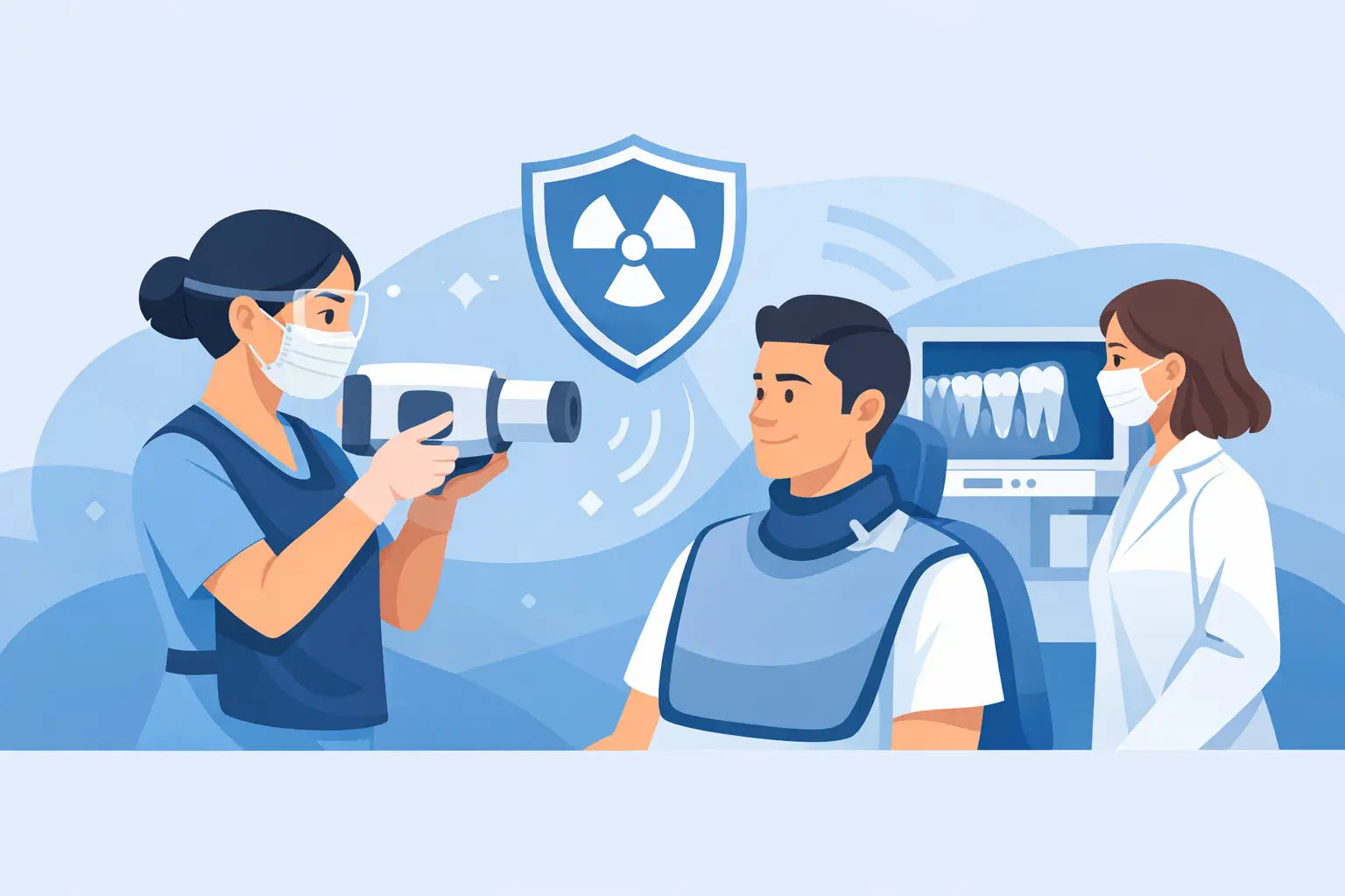
A portable X-ray unit can bring real flexibility to an operatory, a surgical suite, or a mobile-care setting. That flexibility only delivers value when portable radiography safety is built into the clinical routine, not treated as a checklist completed after purchase. For dental teams, the goal is straightforward: obtain diagnostically useful images with the lowest reasonable exposure while protecting patients, operators, and everyone nearby.
Handheld and portable systems are not simply smaller versions of wall-mounted equipment. Their workflow, handling requirements, and scatter-radiation controls demand deliberate training. A well-selected FDA-cleared unit, used according to its instructions for use and applicable state requirements, can support efficient imaging without compromising clinical discipline.
Why Portable Radiography Safety Requires Its Own Workflow
The practical advantage of portable radiography is clear. The operator can bring the unit to the patient instead of moving a patient with limited mobility, interrupting a surgical procedure, or relying on a fixed operatory setup. This is especially useful in mobile dentistry, nursing-facility care, oral surgery, and practices with high-throughput workflows.
The trade-off is that the operator is closer to the source than with many fixed systems. Portable units are engineered with safety features such as backscatter shielding, but those features do not replace correct positioning, trained operation, and adherence to manufacturer instructions. The safest protocol is not based on habit or convenience. It is based on the specific device, the clinical setting, and the rules that apply in the state where the equipment is used.
A portable X-ray unit should be part of the practice radiation-safety program from day one. That means documenting who may operate it, where it may be used, how images are prescribed, and what steps the team follows before every exposure.
Start With Justification and Image Optimization
Radiation safety begins before the unit is turned on. Every exposure should have a diagnostic purpose supported by the patient examination and the clinician's judgment. Taking an image because it is routine, convenient, or likely to be requested later adds exposure without adding clinical value.
Once an image is justified, optimize the technique. Select the correct patient size setting, exposure parameters, receptor, and projection for the diagnostic task. Digital sensors can help reduce retakes when paired with accurate exposure settings and consistent positioning, but they do not make retakes harmless. A clipped apex, overlapped contact, or cone cut still costs time and may require another exposure.
Receptor holders deserve attention here. A stable holder improves alignment and keeps the operator's hands away from the primary beam. Patients should never be asked to hold the sensor or image receptor in place, and staff should not hold it for them. If positioning is difficult because of anatomy, gagging, limited opening, or surgical conditions, pause and adjust the approach rather than improvising during exposure.
Operator Positioning Is a Core Safety Control
For portable radiography safety, the operator's position during exposure is not optional. Follow the device manufacturer's instructions for use precisely, including how to hold the unit, where to stand, and how to use its protective backscatter shield. Do not alter, remove, cover, or defeat a factory-installed safety feature.
Before exposure, verify that no other person is in the immediate area unless their presence is clinically necessary. Patients who require assistance should be managed according to the practice's written protocol and applicable regulations. When a caregiver must remain in the room, that person should receive appropriate instruction and protection and should not be positioned in the path of the primary beam.
Staff should also avoid placing any part of their body in the primary beam. Never hold the tube head, sensor, or patient during an exposure. These rules may seem basic, but they matter most when a schedule is busy, a patient is anxious, or imaging occurs outside the predictable layout of a standard operatory.
Use the Right Room and the Right Boundaries
Portable does not mean unrestricted. A practice should define approved locations for handheld imaging and consider occupancy, traffic flow, nearby workstations, and the presence of nonessential personnel. In a crowded clinical area, the most efficient choice may be to use a designated imaging space rather than exposing beside an open hallway or active treatment bay.
State and local requirements can differ on registration, shielding evaluations, operator credentials, signage, inspections, and documentation. Practice owners should confirm the rules with their state radiation-control authority and follow any direction from their qualified expert or radiation-safety adviser. A protocol that works in one state or facility may not satisfy requirements in another.
Train for the Device, Not Just for Dental X-Rays
A licensed dental professional may be experienced with conventional intraoral imaging and still need device-specific training for a portable system. Training should cover the unit's controls, exposure selection, battery handling, protective shield placement, error messages, charging requirements, cleaning method, and emergency steps.
Teams also need to know when not to use the unit. A damaged shield, cracked housing, loose component, inconsistent exposure, battery fault, or error code should take the device out of service until it is evaluated. Continuing to use equipment that appears damaged creates unnecessary risk and can turn a small service issue into a compliance problem.
Document initial training and refresh it whenever a new operator joins, a device is replaced, or the manufacturer updates its instructions. Brief competency checks are useful because they reveal workflow drift early. If different clinicians use different exposure techniques or positioning habits, the practice can correct the issue before retakes and inconsistent image quality become routine.
Build Safety Into Daily Setup and Maintenance
Reliable imaging depends on equipment condition as much as operator technique. Begin each day with a quick visual inspection of the portable unit, shield, hand grip, controls, charging contacts, and any accessories. Confirm that the device has adequate battery power and that the intended digital receptor, holder, and infection-control barriers are ready before the patient is seated.
Infection control and radiation safety intersect often. Use barriers and cleaning products approved by the manufacturer. Excess liquid, harsh chemicals, or improperly fitted barriers can damage controls and surfaces over time. A damaged unit is not only a service concern; it may affect the practice's ability to maintain a controlled, predictable imaging process.
Keep service records, quality-control documentation, and any required inspection reports organized and easy to retrieve. Monitor retake patterns as well. A rising retake rate can point to technique errors, sensor issues, software settings, or equipment performance concerns. It is a useful quality indicator because it connects patient exposure, clinical efficiency, and staff training in one measurable signal.
Patient Communication Builds Confidence
Patients may be more curious about a handheld X-ray device than a fixed wall unit. A concise explanation helps: the image is being taken because it supports diagnosis or treatment planning, the team uses modern digital imaging protocols, and the device is operated under established safety procedures.
Avoid promising that an X-ray has no radiation. A better approach is to explain that dental radiography uses low exposure levels and that the practice limits exposures to those that are clinically necessary. This is honest, calm, and consistent with professional standards.
Protective apparel policies should also be consistent with current regulations, professional guidance, and the practice's own protocol. The appropriate use of thyroid collars, aprons, and other protective measures can vary by patient, procedure, equipment, and jurisdiction. The key is to avoid a one-size-fits-all response and ensure the entire team follows the same evidence-informed process.
Choose Equipment With Support Behind It
Price matters when investing in imaging technology, but portable X-ray purchasing should not be reduced to price alone. Evaluate FDA clearance, manufacturer reputation, included safety features, service availability, warranty terms, training resources, and access to replacement components. The least expensive unit can become the costliest option if support is difficult to reach or documentation is incomplete.
For practices expanding into mobile care or upgrading digital workflows, the right supplier should help clarify practical questions before the purchase: What training is required? What accessories support correct positioning? How will the device fit into the existing sensor and software workflow? What service path is available if a problem occurs? ProElite Dental Supply approaches equipment selection with that clinical and operational perspective in mind.
A portable X-ray unit earns its place in a practice when it makes care more flexible without making safety harder to manage. Give the team a clear protocol, dependable equipment, and permission to slow down when a setup is not right. That is how efficient imaging remains safe, repeatable, and worthy of patient trust.