

When Should Dentists Use CBCT in Practice?
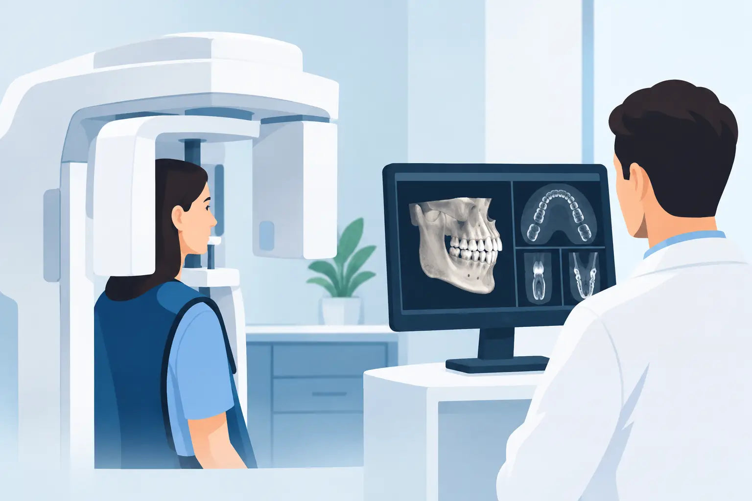
A two-dimensional image can answer many clinical questions. But when anatomy overlaps, a root canal disappears behind cortical bone, or an implant site demands a clearer safety margin, guessing is not a treatment plan. Knowing when should dentists use CBCT helps practices obtain the diagnostic detail they need without making three-dimensional imaging a default for every patient.
CBCT is most valuable when conventional radiographs cannot adequately answer a specific diagnostic or treatment-planning question. Its strength is spatial information: the buccolingual dimension, relationship to vital structures, extent of osseous pathology, tooth position, and hard-tissue anatomy that may be obscured on periapical or panoramic imaging.
The goal is not simply to produce a more impressive image. It is to make a more defensible clinical decision, using the smallest appropriate field of view and imaging settings for the case.
When Should Dentists Use CBCT for Diagnosis?
A practical rule is straightforward: use CBCT when the anticipated diagnostic benefit is likely to change or confirm a treatment decision and lower-dose two-dimensional imaging is insufficient.
That distinction matters. CBCT should not replace a clinical examination, periodontal charting, conventional radiographs, or sound judgment. For routine caries detection, standard periodontal evaluation, and many straightforward restorative cases, intraoral imaging remains the efficient choice. It is familiar, fast, and typically exposes the patient to less radiation.
CBCT becomes a stronger option when the clinical picture and 2D images do not agree, when anatomy is complex, or when treatment carries meaningful risk. In these situations, a well-selected scan can prevent surprises that cost time, compromise outcomes, or require an avoidable referral.
Implant planning and site evaluation
Implant dentistry is one of the clearest indications for CBCT. A three-dimensional scan helps the clinician assess ridge width and height, bone contours, sinus anatomy, the inferior alveolar canal, mental foramen, incisive canal, and adjacent root positions. It also supports virtual implant positioning based on restorative objectives rather than placing an implant into an incomplete two-dimensional picture.
This does not mean every edentulous space requires the largest available scan. Select the field of view that captures the planned site and critical adjacent anatomy. A limited field is often appropriate for a single-tooth site, while full-arch rehabilitation, multiple implants, or complex grafting may justify a larger volume.
For practices investing in guided surgery or digital workflows, CBCT can also provide the imaging foundation for merging scan data and planning surgical guides. The return is not just in technology adoption. It is in improved communication, more predictable surgical preparation, and a clearer record of the rationale behind treatment.
Complex endodontic diagnosis and retreatment
Endodontic cases can look deceptively simple on a periapical image. CBCT may be warranted when persistent symptoms, an equivocal lesion, suspected root fracture, unusual anatomy, resorption, perforation, or a missed canal would alter the treatment plan.
For example, a mandibular molar with recurring symptoms after treatment may require more than another angled radiograph. A limited-field CBCT scan can help reveal untreated anatomy, the relationship of a lesion to roots, or evidence of external root resorption. In retreatment planning, it may also help assess posts, separated instruments, previous obturation, and proximity to critical structures.
The trade-off is image interpretation. Small fields and high-resolution protocols can offer excellent detail, but artifacts from restorations and endodontic materials may still obscure findings. CBCT should support the endodontic diagnosis, not be treated as a stand-alone answer.
Impacted teeth and surgical anatomy
Impacted canines, third molars, supernumerary teeth, and teeth displaced by trauma are common reasons to consider CBCT. The key question is whether knowing the exact three-dimensional relationship will influence the surgical approach, risk discussion, or referral decision.
For impacted maxillary canines, CBCT can clarify the tooth's position relative to adjacent roots and help identify possible resorption. For mandibular third molars, it may be appropriate when panoramic findings suggest close proximity to the inferior alveolar canal and the information could change the surgical plan.
Oral surgery cases involving retained root fragments, complex extractions, suspected sinus communication, or bony pathology may also benefit from volumetric imaging. The value is strongest when the scan narrows uncertainty before the procedure, rather than documenting information that will not affect care.
Trauma, pathology, and unexplained findings
CBCT can be particularly useful for suspected facial fractures, dentoalveolar trauma, root fractures, and localization of foreign objects. It may also help characterize the boundaries and effects of osseous lesions, evaluate cortical expansion or perforation, and guide referral when a finding exceeds the scope of general practice.
A scan is not automatically indicated for every radiolucency or traumatic event. Consider the patient’s symptoms, the quality of available images, the urgency of the condition, and whether the result will affect immediate management. If a lesion or finding requires specialist interpretation, referral should be part of the plan, not an afterthought.
When Should Dentists Use CBCT in Orthodontic and Airway Cases?
CBCT has a role in selected orthodontic cases, especially impacted teeth, skeletal asymmetry, craniofacial concerns, root position questions, and treatment planning where conventional images do not provide enough information. It should not be used as routine imaging for every orthodontic patient simply because a 3D model is available.
Airway evaluation requires similar restraint. CBCT can visualize airway anatomy, but a static scan does not independently diagnose sleep-disordered breathing or replace a medical evaluation. If airway concerns arise during dental assessment, imaging findings should be integrated with clinical screening and appropriate medical or sleep-specialist referral.
For both categories, clear documentation matters. Record the clinical indication, why 2D imaging was inadequate, the selected field of view, and how the findings influenced care. This supports quality assurance and helps the practice maintain a disciplined, patient-centered imaging protocol.
Choose the Right Scan, Not Just a CBCT Scan
Clinical value depends on more than owning a CBCT unit. Practices need an imaging workflow that matches their case mix, operator training, space, and referral relationships.
Start with field of view. A small field can provide targeted detail for endodontics, an implant site, or a localized surgical question. Medium and large fields may suit multi-site implant planning, full-arch assessment, orthodontic evaluation, or broader pathology review. Larger is not inherently better. It can mean more anatomy to interpret, more incidental findings to manage, and potentially greater patient exposure.
Resolution also involves trade-offs. High-resolution protocols can be useful for fine endodontic or root-fracture questions, but may increase scan time, noise, or dose depending on the system and settings. Select protocols based on the diagnostic task, not a one-size-fits-all preset.
Interpretation is equally important. The clinician who orders the scan is responsible for reviewing the entire acquired volume, not only the region of immediate interest. Establish a reliable process for reviewing studies and obtaining interpretation support when needed. This is especially important for incidental findings outside the planned treatment area.
Building a CBCT Workflow That Supports ROI
For a practice owner, the decision to add CBCT is both clinical and operational. The best investment is one that fits real patient demand. Implant-focused offices, endodontic practices, oral surgery providers, and multidisciplinary groups may see frequent, direct use. A general practice may benefit when it performs enough implants, complex extractions, endodontic diagnosis, and treatment planning to reduce outside referrals and improve case acceptance.
Before purchasing, evaluate more than the unit price. Consider available fields of view, software usability, sensor technology, training, installation requirements, warranty coverage, service response, and ongoing support. FDA-cleared equipment, dependable supplier communication, and a clear training plan protect the practice long after the delivery date.
ProElite Dental Supply works with licensed dental professionals looking for advanced imaging capability at smart-practice pricing. For buyers comparing systems, the productive question is not which unit has the longest feature list. It is which system gives the practice the right diagnostic range, workflow efficiency, and support for the cases it treats every week.
A disciplined CBCT protocol makes advanced imaging feel less like a capital expense and more like a clinical advantage. Use it when the third dimension meaningfully changes the decision, keep the scan focused on the question at hand, and let every image earn its place in the patient’s care.