

What Radiation Shielding Is Required for Dentistry?
A new X-ray unit can improve diagnostic workflow on day one. A room that has not been evaluated for radiation protection can delay installation, trigger costly changes, and create avoidable compliance questions. That is why practices asking what radiation shielding is required need an answer tied to their actual equipment, room layout, patient volume, and state requirements - not a one-size-fits-all wall specification.
For most dental practices, the goal is straightforward: keep exposure to patients, staff, and the public as low as reasonably achievable while allowing efficient imaging. The details, however, change substantially between intraoral radiography, panoramic imaging, CBCT, and portable X-ray systems.
What radiation shielding is required in a dental office?
Radiation shielding is required when a room's design, nearby occupied areas, and projected radiation workload show that barriers are needed to keep exposure outside the room within applicable limits. In the United States, dental X-ray installations are governed through a combination of federal equipment standards, state radiation-control rules, local building requirements, and professional radiation-safety guidance.
The practical answer is often one of three designs: a protected operator position behind a fixed barrier, structural shielding built into walls or other barriers, or a layout that allows the operator to stand at a safe distance and angle from the primary beam. Which approach is acceptable depends on the installation and the rules in your state.
A shielding plan should account for the type of system, its technical exposure factors, beam direction, anticipated number of exposures, room dimensions, wall construction, adjacent rooms, and occupancy on the other side of each barrier. A storage closet presents a different risk profile than a neighboring operatory, waiting room, apartment, or office occupied all day.
Start with the imaging system, not a standard wall recipe
Dental equipment does not create the same shielding demand across every modality. Treating all X-ray rooms alike can lead to overspending on construction or, worse, installing insufficient protection.
Intraoral X-ray rooms
Conventional wall-mounted intraoral units generally have a relatively focused beam and short exposure times. In many existing operatories, a properly located protective barrier or control position may be sufficient. Whether lead-lined walls are necessary depends on the room's construction, the location of adjacent occupied spaces, and your state’s requirements.
The operator should not hold the receptor, tube head, or patient during exposure except in rare, permitted clinical circumstances. A protected position is commonly arranged so the operator remains out of the primary beam and away from scatter radiation, typically at an appropriate distance and angle to the beam path. A fixed barrier with a leaded viewing window may be the cleanest operational solution in a busy practice.
Panoramic and cephalometric systems
Panoramic imaging usually involves a lower shielding burden than many clinicians expect because the beam is narrow and moves around the patient. Still, the equipment should be placed so the beam is not directed toward routinely occupied areas without an appropriate barrier assessment.
A panoramic room may work with standard construction and an operator position outside the room or behind a barrier, but that conclusion should follow a review of the specific unit and layout. Do not assume an open-area installation is acceptable simply because a prior panoramic unit occupied the space.
CBCT systems
CBCT calls for the most careful planning in many dental offices. Field of view, exposure settings, scan protocols, use frequency, and the position of occupied spaces can materially affect the shielding design. A small-field endodontic CBCT used selectively has a different workload from a large-field system supporting implants, oral surgery, orthodontics, and high-volume imaging.
For CBCT, a formal shielding evaluation is often the smart investment before signing off on construction. It can determine whether existing gypsum-board partitions are adequate, where supplemental lead or other shielding belongs, and whether an alternate room layout reduces cost without compromising protection.
Portable dental X-ray units
Portable X-ray units can support mobile care, nursing-home dentistry, hospital rounds, and flexible practice workflows. They do not eliminate radiation-safety responsibilities. The unit must be FDA-cleared for its intended use, operated according to manufacturer instructions, and used in a manner permitted by state regulations.
Portable use requires special attention to operator positioning, bystander control, patient-area selection, and exposure documentation. The device’s backscatter shield is part of its engineered safety design, but it is not a substitute for training, distance, sound technique, and compliance with applicable state rules. In a fixed dental office, a portable unit should not be treated as a shortcut around a thoughtful radiation-safety program.
Primary barriers, secondary barriers, and protected positions
Shielding calculations distinguish between primary and secondary barriers. A primary barrier is placed where the useful X-ray beam could directly strike. A secondary barrier protects against scatter from the patient and leakage radiation from the X-ray tube housing.
This distinction matters because the primary beam requires considerably more protection. Thoughtful equipment placement can often keep the primary beam aimed toward a less-sensitive direction, such as an exterior wall or an area with limited occupancy. That planning decision may reduce the amount of structural shielding required.
Barrier materials may include lead sheet, lead-lined drywall, leaded glass, concrete, masonry, or other materials with documented equivalent protective value. The correct material and thickness should be specified in lead equivalency or another recognized calculation method, not selected by guesswork. Door frames, doors, windows, pass-throughs, wall penetrations, and ceiling or floor paths all deserve review. A well-shielded wall can still fail the intended design if a gap around a door or utility opening is overlooked.
Why state rules and occupancy matter
There is no universal answer such as “every dental X-ray room needs one-sixteenth-inch lead.” Some jurisdictions prescribe specific construction or registration expectations. Others rely more heavily on documented shielding calculations and inspections. Existing facilities, tenant improvements, and new construction can also be handled differently under local rules.
Occupancy is equally significant. The wall between an X-ray room and a hallway used briefly by staff may need a different design than the wall bordering a pediatric operatory, shared medical suite, or public waiting area. Upper and lower floors matter, too. If another tenant occupies the space above a CBCT room, the ceiling assembly may need evaluation even when every side wall appears satisfactory.
Before construction begins, contact the state radiation-control program or the qualified expert supporting your project. Your installer, equipment manufacturer, architect, and contractor should work from the same room plan and shielding assumptions. Late changes in beam orientation, room use, or machine model can invalidate an otherwise sound plan.
Use a qualified shielding assessment when the risk is higher
A qualified medical physicist, health physicist, or other state-accepted radiation-safety professional can prepare a shielding design report using the equipment specifications and architectural drawings. This is especially valuable for CBCT, unusual room layouts, multi-tenant buildings, imaging centers, and practices with high projected volume.
The report commonly identifies each barrier, its required protective equivalency, the expected occupancy beyond it, and any installation notes. Keep it with the practice’s radiation-safety records. It provides a defensible basis for construction decisions and makes future inspections, equipment replacement, and facility expansion far easier to manage.
A post-installation survey may also be required or advisable. This verifies that barriers were installed as designed and that the system, room, and operator location function as intended. It is far less expensive to confirm protection before a room is fully finished and in daily use than to reopen walls later.
Shielding is only one part of radiation safety
Physical barriers work best within a disciplined imaging program. Practices should use current equipment, maintain it according to manufacturer guidance, select the smallest clinically appropriate field of view, and avoid repeat exposures through proper positioning and staff training. Digital sensors and optimized imaging workflows can support diagnostic quality while helping reduce unnecessary retakes.
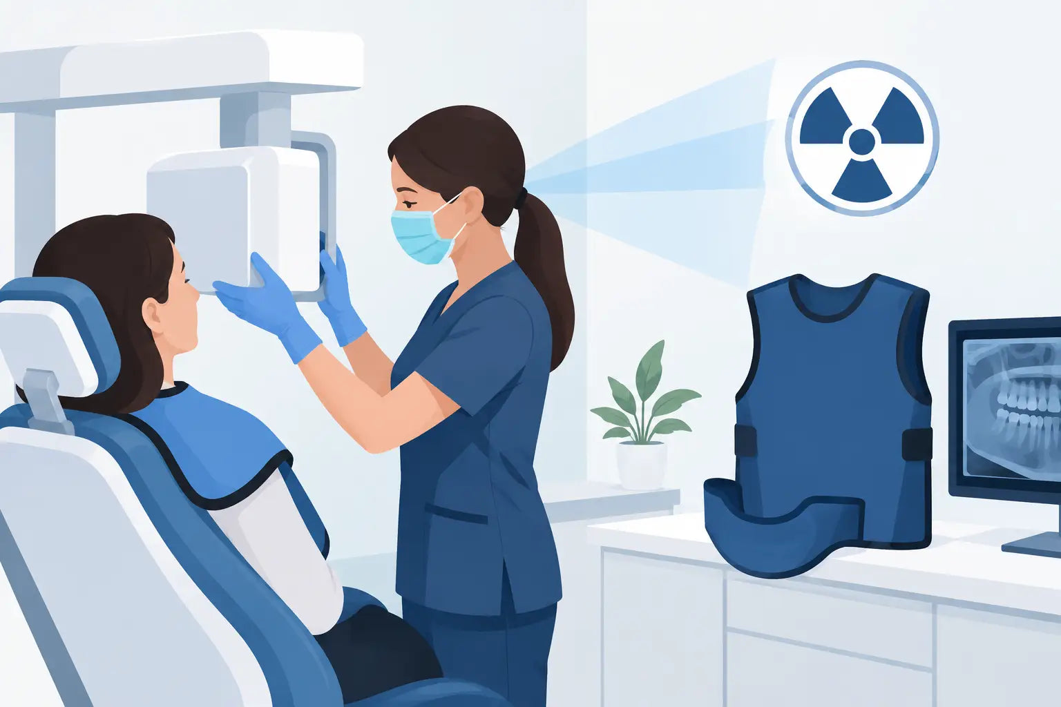
Patient lead aprons and thyroid collars should be used according to current clinical guidance, equipment instructions, and state requirements. They are not a replacement for room shielding or sound exposure technique. Staff personal monitoring may be required in certain settings or recommended based on workload and state policy, but it should be determined as part of the practice’s broader radiation-safety program.
When purchasing new imaging technology, ask for the technical documentation early - before finalizing the room. ProElite Dental Supply helps practices evaluate professional imaging options with an eye toward clinical performance, workflow, and practical implementation. The right system is not just one that fits the budget; it is one your facility can install, operate, and support with confidence.
A well-planned X-ray room protects more than compliance. It protects your team’s time, your construction budget, and the confidence patients place in your practice every time imaging is part of care.