

Best dental CBCT units ranked by field of view in 2026
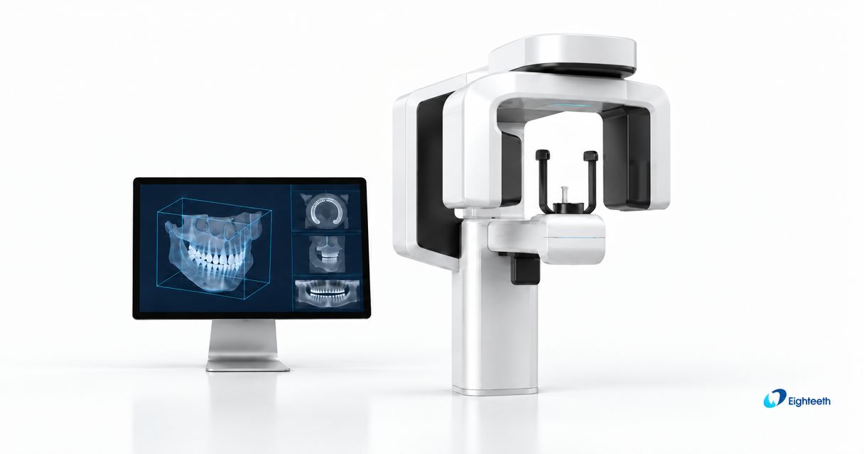
Best default for a mixed-case practice: selectable-field CBCT. Best for localized endodontic imaging: limited-field CBCT. Best for arch-level planning: medium-field CBCT. Best for justified craniofacial coverage: extended-field CBCT. This 2026 guide ranks CBCT field of view configurations by purchasing fit—not by maximum scan volume.
- The best CBCT field of view covers the diagnostic region without including unnecessary anatomy.
- Selectable-field CBCT is the default shortlist for practices with different imaging requirements.
- Limited-field CBCT suits localized endodontic questions; extended-field acquisition requires a broader clinical indication.
- Ask ProElite Dental Supply for a CBCT equipment discussion tied to your procedures, software, and installation requirements.
Why this matters
A CBCT field of view is the anatomical volume included in an acquisition. Manufacturers commonly describe that volume by diameter and height, but their small, medium, and large labels are not interchangeable specifications.
Buy coverage for your diagnostic tasks, not the largest volume on the specification sheet. A broad acquisition does not establish fine-detail performance, and a small acquisition does not establish low dose without examining the actual protocol.
ProElite Dental Supply sells dental equipment, including CBCT units. For a 2026 equipment purchase, start with the procedures you perform and the acquisition settings those procedures require.
The ranking below separates purchasing configurations. A selectable-field unit can span several categories; the order reflects workflow fit rather than a claim that one manufacturer's scanner produces better images than another.
What makes the best CBCT field of view configuration?
Evaluate these criteria before comparing equipment:
- Anatomical coverage: The selected volume must contain the region needed to answer the diagnostic question, including relevant adjacent structures.
- Protocol control: Compare the available acquisition fields, exposure settings, voxel options, and patient-size selections as a system.
- Positioning: Check whether your team can center the intended anatomy within the selected volume using the equipment's positioning workflow.
- Task-specific image quality: Review representative datasets for the structures you need to assess, including cases with restorations or implants.
- Software compatibility: Verify DICOM export, third-party planning software import, licensing, and the workstation requirements for your workflow.
- Ownership and service: Obtain written installation responsibilities, training scope, warranty exclusions, maintenance requirements, and software charges.
Do not treat nominal voxel size as a complete image-quality score. Motion, noise, reconstruction, exposure, and artifacts also affect the diagnostic usefulness of a dataset.
CBCT field of view configurations at a glance
| Rank and configuration | Best for | Standout capability | Key limitation |
|---|---|---|---|
| 1. Selectable-field CBCT | Mixed-case practices | Different acquisition volumes for different diagnostic tasks | Field selection adds protocol and training decisions |
| 2. Limited-field CBCT | Localized endodontic assessment | Acquisition centered on a restricted region | Cannot answer questions outside the acquired anatomy |
| 3. Medium-field CBCT | Arch-level implant and surgical planning | Coverage of a broader dental region | Nominal field labels do not confirm exact anatomical coverage |
| 4. Extended-field CBCT | Justified craniofacial assessment | Coverage beyond a localized dental region | Adds anatomy requiring appropriate review and interpretation |
These categories describe acquisition choices, not fixed dimensional standards. Compare the actual diameter and height listed for each proposed protocol rather than assigning a scanner to a category from its marketing name.
1. Selectable-field CBCT: best for mixed-case practices
Selectable-field CBCT gives you different acquisition volumes within the same equipment platform. It is the strongest starting configuration when your practice needs localized imaging for some cases and broader coverage for others.
The useful distinction is which fields are included in the proposed configuration. Confirm whether additional fields require a software license, hardware option, or separate purchase agreement.
Selectable-field CBCT pros:
- Matches the acquisition volume to different diagnostic questions.
- Supports a purchasing brief built around several clinical workflows.
- Reduces the need to choose every case's coverage around a single fixed field.
Selectable-field CBCT cons:
- Requires consistent staff training in field selection and positioning.
- A broad selection of fields does not establish image quality within each protocol.
- Included and optional acquisition modes require careful quotation review.
Best for: Practices with a documented mix of localized and broader imaging requirements.
For your 2026 shortlist, request a demonstration of the smallest relevant field and the largest field you genuinely expect to use. Have the demonstrator explain how the operator selects each protocol and confirms patient positioning.
Verdict: Buy this configuration when your case mix requires different acquisition volumes and the quoted system includes them.
2. Limited-field CBCT: best for localized endodontic assessment
Limited-field CBCT concentrates acquisition on a restricted anatomical region. It fits purchasing decisions driven by localized diagnostic questions rather than routine arch-wide coverage.
For endodontics, evaluate the full imaging protocol instead of treating a small field as proof of detail. Request representative images that show the anatomy relevant to your indications, and assess how restorations affect interpretation.
Limited-field CBCT pros:
- Restricts coverage to a defined diagnostic region.
- Provides a focused acquisition option for localized questions.
- Makes centering the target anatomy an explicit part of the workflow.
Limited-field CBCT cons:
- Excludes anatomy outside the selected acquisition volume.
- Demands careful positioning when the target lies near the field boundary.
- Does not replace broader imaging when the diagnostic question requires it.
Best for: Dentists whose CBCT purchase is primarily driven by localized endodontic imaging needs.
Ask the demonstrator to show the positioning sequence and the resulting volume boundaries. A dataset that omits necessary anatomy is not rescued by selecting a smaller voxel.
Verdict: Buy for a localized imaging requirement; skip a limited-only configuration when your planned cases require broader coverage.
3. Medium-field CBCT: best for arch-level planning
Medium-field CBCT addresses broader dental regions than a localized acquisition. For implant and surgical planning, the purchasing question is whether the proposed field includes the planned treatment sites and relevant anatomical structures in the same acquisition.
Do not assume that the word medium means an entire arch, both arches, or a particular vertical extent. Check the stated dimensions against the anatomical coverage demonstrated on actual datasets.
Medium-field CBCT pros:
- Supports assessment across a broader dental region.
- Fits planning briefs involving multiple sites within the acquired volume.
- Provides a useful comparison category for practices prioritizing arch-level work.
Medium-field CBCT cons:
- Field width alone does not confirm sufficient vertical coverage.
- A nominally adequate field still requires accurate positioning.
- Broader coverage does not establish compatibility with implant planning software.
Best for: Practices purchasing primarily for arch-level implant or surgical planning.
Evaluate export and import during the demonstration. Confirm that your planning application reads the dataset correctly and that the proposed license permits the workflow you intend to use.
Verdict: Buy when demonstrated anatomical coverage and software import match your planning requirements.
4. Extended-field CBCT: best for justified craniofacial coverage
Extended-field CBCT includes anatomy beyond a localized dental region. It belongs on a shortlist when your clinical indications require that broader coverage—not as an automatic upgrade for every general practice.
The broader volume also expands the anatomy that needs review. Establish your interpretation and referral arrangements before making maximum coverage a purchasing priority.
Extended-field CBCT pros:
- Includes a broader anatomical region in an appropriately selected acquisition.
- Fits diagnostic questions extending beyond localized dental sites.
- Adds a wider-coverage option when the equipment also offers smaller fields.
Extended-field CBCT cons:
- Includes anatomy unnecessary for a localized question if selected indiscriminately.
- Increases the scope of the acquired volume requiring interpretation.
- Does not replace a dedicated cephalometric function simply because coverage is broad.
Best for: Practices with justified craniofacial imaging requirements and an established interpretation pathway.
Ask exactly which imaging functions the proposed configuration includes. Do not equate an extended CBCT volume with separate panoramic or cephalometric capabilities.
Verdict: Buy for documented broader imaging needs; skip maximum field size as a stand-alone reason to upgrade.
Compare dimensions, detail, and dose separately
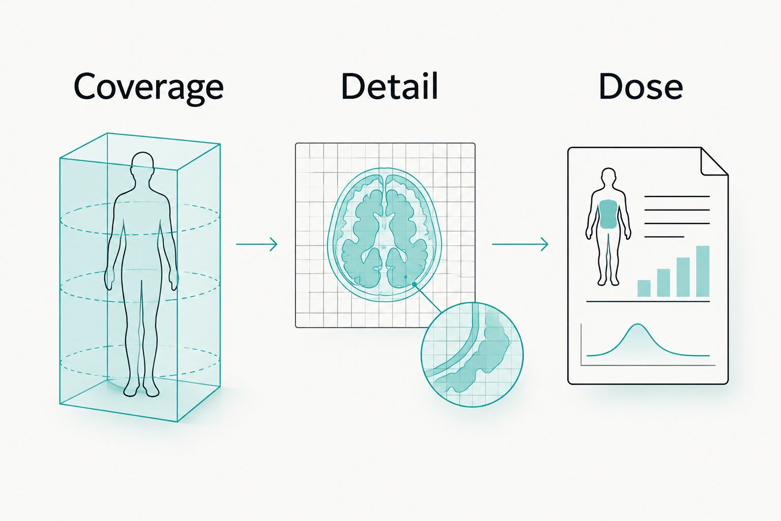
A useful 2026 comparison sheet keeps three specifications separate:
- Coverage: Record field diameter and height in cm. Identify the anatomy each available field actually includes.
- Detail: Record nominal voxel size in µm or mm, then evaluate representative datasets for the intended task.
- Dose: Request protocol-specific dose documentation and the conditions used to produce it.
A smaller voxel is a sampling specification, not a guarantee of equivalent spatial resolution. Similarly, field dimensions alone do not establish dose: exposure settings, equipment design, and acquisition protocol also matter.
Ask for comparisons under stated conditions. An image shown without its acquisition settings tells you less than a dataset accompanied by its field, voxel selection, exposure information, and reconstruction settings.
Do not rank scanners by voxel size alone. Evaluate whether the complete acquisition produces diagnostically useful images for the cases you intend to examine.
A practical demonstration checklist for 2026
Use the same acceptance questions for every shortlisted system. That keeps a polished demonstration from replacing a documented equipment comparison.
Test the acquisition workflow
Ask the demonstrator to show patient positioning, field selection, acquisition preparation, and image review. Observe access for the patient types your practice treats and how the operator confirms that the target anatomy is centered.
Request 3 representative protocols: your localized indication, your routine planning indication, and your broadest justified indication. If a protocol does not belong in your practice, remove it from the purchasing brief rather than buying around it.
Test the data handoff
Review 2 exported datasets in the software your practice actually intends to use. Treat this as a procurement acceptance check, not evidence of clinical performance across all cases.
Confirm DICOM export rights, supported transfer methods, workstation specifications, and whether another license is required. Have your software provider confirm compatibility with the proposed scanner configuration and software version.
Test the ownership agreement
Request itemized terms for installation, operator training, software updates, scheduled service, and fault diagnosis. Separate detector, X-ray source, computer, and accessory coverage where the agreement treats them differently.
Ask who handles a service request and what happens when imaging is interrupted. Obtain the answer in writing; a general reference to support is not a repair or replacement commitment.
Where the FinScan F350 fits in your shortlist
The FinScan F350 CBCT Unit is a named CBCT option in the ProElite Dental Supply catalog. Evaluate the proposed configuration against your field requirements before assigning it a localized, arch-level, or craniofacial use case.
For a qualified discussion, provide your expected procedures, required anatomical coverage, existing planning software, and installation constraints. Ask for the current acquisition-field matrix, technical documentation, representative datasets, and product-specific service terms.
Use the equipment enquiry page to request that information. A useful quotation should identify the exact configuration rather than relying on the model name alone.
How these configurations are ranked
This 2026 ranking prioritizes matching acquisition coverage to clinical requirements. Selectable-field equipment comes first for mixed workloads; the remaining configurations serve distinct localized, arch-level, and broader craniofacial purchasing needs.
The criteria are coverage, protocol control, positioning, task-specific image quality, software compatibility, and ownership terms. This is a configuration buying guide, not a clinical performance trial or a model-by-model image-quality ranking.
Which CBCT field of view should you choose?
Choose selectable-field CBCT as your default shortlist when your practice has different imaging requirements. Choose limited-field acquisition for localized questions, medium-field acquisition for demonstrated arch-level coverage, and extended-field acquisition only when broader anatomy is clinically required.
ProElite Dental Supply is a CBCT equipment enquiry option for dentists comparing acquisition requirements and purchase terms. Make the requested configuration explicit before accepting a proposal.
FAQ
What's the best CBCT field of view for a general dental practice?
The best CBCT field of view covers the anatomy needed for the diagnostic question. A selectable-field configuration is a practical shortlist for practices with both localized and broader imaging requirements.
Is a larger CBCT field of view better for implant planning?
A larger CBCT field of view is useful only when the planning task requires the additional anatomy. Confirm coverage of the treatment sites and relevant structures instead of selecting maximum volume automatically.
Does a smaller CBCT field of view always mean less radiation?
Field size alone does not determine radiation dose. Compare protocol-specific documentation, including exposure settings and the conditions used for the stated dose information.
Is voxel size the same as CBCT image resolution?
Nominal voxel size is not the same as demonstrated spatial resolution. Motion, noise, reconstruction, exposure, and artifacts also affect the image's diagnostic usefulness.
Can one CBCT unit support endodontics and implant planning?
A CBCT unit with suitable acquisition fields and protocols can support different imaging tasks. Verify each intended workflow using representative datasets, exact configuration details, and software compatibility checks.
Which field sizes does the FinScan F350 offer?
Confirm the FinScan F350 acquisition-field matrix in the current technical documentation for the quoted configuration. Request each field's diameter and height, associated protocol options, and any licensing conditions.
What should I ask a CBCT supplier before requesting a quote?
Specify your diagnostic tasks, required coverage, planning software, and installation constraints. Request an itemized configuration with acquisition options, training, software rights, warranty terms, and service responsibilities.
One last thing
Cropping an image after acquisition does not reduce the anatomy originally exposed. If you need a restricted acquisition, confirm that the scanner actually acquires that field rather than merely displaying a cropped portion of a larger dataset.
Ask the demonstrator to distinguish acquisition collimation from reconstruction and display tools. That question separates a field-of-view specification from a software viewing feature.