

Dental Sensor Resolution: What Dentists Need
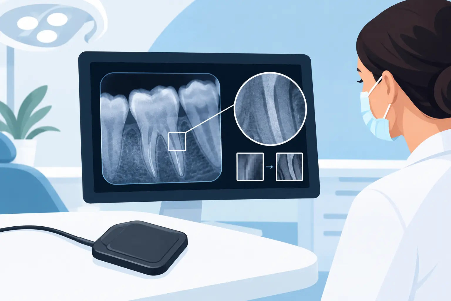
A periapical image can look impressively sharp on a monitor and still fail to answer the clinical question. When evaluating dental sensor resolution, the real concern is not simply whether a product lists a high number. It is whether the sensor consistently helps clinicians see caries margins, periodontal bone levels, root anatomy, apical changes, and endodontic files at the exposure levels and workflow speed the practice needs.
Resolution matters. It is not the only imaging specification that matters, and it is often misunderstood in purchasing conversations. A smarter comparison looks at how resolution works alongside pixel size, contrast performance, software processing, sensor durability, ergonomics, and the quality of the complete imaging system.
What Dental Sensor Resolution Really Measures
Dental sensor resolution describes how well an intraoral sensor can distinguish small, closely spaced structures. It is commonly expressed in line pairs per millimeter, written as lp/mm. A line pair is one dark line and one light line. The higher the number of line pairs a system can distinguish, the finer the detail it can theoretically reproduce.
That definition is useful, but it has limits. A published lp/mm specification may represent a sensor's theoretical capability under controlled test conditions. It does not automatically predict how well a radiograph will perform in a real operatory, with patient movement, variable angulation, positioning challenges, exposure differences, and software enhancement applied to the final image.
Pixel size is closely related. Smaller pixels can sample finer detail, which may support higher spatial resolution. However, smaller pixels can also collect less signal. If a sensor produces insufficient signal at practical exposure settings, image noise can offset the benefit of a finer pixel grid. The best result is not the highest number on a specification sheet. It is diagnostically useful detail with dependable image quality.
Spatial Resolution Is Only One Part of Image Quality
Spatial resolution answers one question: how finely can the sensor separate adjacent details? Diagnostic imaging asks several others. Can the system differentiate subtle shades of gray in low-contrast areas? Does the image remain clear without excessive grain? Can the clinician view the anatomy at an appropriate scale without artificial-looking processing? Does the image appear quickly and reliably in the practice management or imaging workflow?
Contrast resolution is especially relevant when evaluating early carious changes, periodontal structures, and subtle periapical findings. A sensor with strong contrast performance may provide more practical diagnostic value than one that advertises a marginally higher lp/mm rating but produces less balanced images.
Noise also deserves attention. Noise is the random visual variation that can make an image look mottled or grainy. It can hide fine anatomy and make subtle findings harder to interpret. Signal-to-noise ratio, sensor electronics, exposure technique, and image processing all influence how clean an image appears.
How to Compare Dental Sensor Resolution Specs
When comparing sensors, begin by confirming that the specifications are presented consistently. Manufacturers may use different testing methods, different terminology, or different values for the sensor hardware and the complete imaging chain. An lp/mm figure is worth reviewing, but it should never be the sole buying criterion.
Ask for representative clinical images captured with the same type of procedure you perform every day. For a general practice, that may include bitewings and periapicals. Endodontic practices should evaluate file visibility, canal anatomy, and apical detail. Implant and surgical offices may prioritize predictable bone-level visualization and a sensor size that supports accurate positioning in challenging posterior areas.
Review the image on the monitor configuration used in the office. A high-quality sensor cannot fully compensate for an aging, poorly calibrated display. Likewise, imaging software settings can make a major difference. Over-sharpening may create edge artifacts that look dramatic but do not represent true anatomy. Excessive smoothing can erase fine detail. The goal is an image that looks natural, readable, and repeatable.
Look Beyond a Single lp/mm Number
A practical equipment evaluation should include these connected factors:
- Pixel pitch and active area: Pixel size affects sampling detail, while the active imaging area affects how much anatomy the sensor captures.
- Contrast and noise performance: These influence whether fine structures are visible, not just whether the image appears sharp.
- Exposure latitude: A sensor that delivers usable images across reasonable exposure variation can reduce retakes and simplify daily operation.
- Software compatibility: Confirm the system works reliably with the imaging software, computers, and acquisition workflow in your practice.
- Sensor construction and warranty support: Cables, housings, connectors, and replacement support directly affect long-term ownership cost.
When Higher Resolution Helps Most
Higher effective resolution can be valuable in procedures where small structures and fine margins matter. Endodontic imaging is an obvious example. Clinicians may need clear visualization of canal pathways, file progression, working length references, root fractures, and subtle apical changes. A sensor that renders detail cleanly can support more confident chairside decisions.
Restorative dentistry also benefits when radiographs make it easier to evaluate interproximal contacts, recurrent caries concerns, and restoration margins. In periodontal assessment, the ability to view crestal bone and lamina dura clearly supports consistent charting and treatment planning.
Still, a higher advertised resolution does not mean every practice needs the most aggressive specification available. If the sensor is difficult to position, uncomfortable for patients, slow to acquire, or unreliable after regular use, the theoretical gain can disappear. A fast, durable system that produces consistent diagnostic images may create a stronger return on investment than a more expensive option with only a modest resolution advantage.
Sensor Size, Positioning, and Clinical Reality
Sensor resolution cannot be separated from positioning. Even the best sensor cannot correct for cone cuts, overlapping contacts, motion, poor receptor placement, or incorrect horizontal angulation. Teams should evaluate whether a sensor's size and thickness work well with their patient population and existing positioning system.
Size 1 sensors are commonly useful for smaller mouths, pediatric patients, and certain anterior views. Size 2 sensors are the standard choice for many adult bitewing and periapical applications because they provide a larger active area. The right selection depends on the procedures performed and the comfort needs of the patient base.
A sensor's cable design and housing shape also influence positioning. A stiff cable or bulky housing can make posterior placement more difficult, particularly for patients with limited opening, a sensitive gag reflex, or tori. Those challenges can lead to retakes, which affect time, radiation exposure, and patient experience more than a small difference in published resolution ever will.
Exposure Settings Still Matter
Digital sensors are often more forgiving than film, but they are not exposure-proof. Underexposure may increase visible noise and reduce diagnostic clarity. Overexposure can limit useful information, even if the software adjusts brightness afterward. The software can improve presentation, but it cannot recreate detail that was never captured.
Work with the sensor manufacturer's recommended exposure guidance for your X-ray source, then establish office protocols by receptor size and exam type. Review images during training and calibrate technique before drawing conclusions about sensor performance. A sensor that initially appears soft or noisy may be paired with suboptimal exposure settings, an incompatible X-ray unit, or inconsistent positioning.
For portable X-ray workflows, technique discipline is equally important. Stable positioning, correct source-to-receptor alignment, and appropriate exposure selection help the practice gain the efficiency benefits of portable imaging without sacrificing diagnostic consistency.
Questions to Ask Before You Buy
Before investing in a new intraoral sensor, ask the supplier whether the device is FDA-cleared for its intended use and whether it is compatible with your current software and operating environment. Confirm what is included: acquisition software, drivers, sensor holders, calibration requirements, warranty terms, and support for installation or troubleshooting.
Also ask what happens after the sale. A sensor is a daily-use clinical device, not a one-time consumable. Fast technical response, clear warranty handling, and access to replacement options matter when an imaging issue threatens the schedule. For multi-location groups and high-volume practices, standardizing on a dependable platform can simplify training, support, and inventory planning.
Price should be evaluated in that same practical context. Premium legacy systems are not the only path to professional image quality. Well-supported, FDA-cleared digital imaging equipment can help practices modernize their workflow while keeping capital costs under control. That balance between clinical performance and sensible purchasing is where a supplier relationship earns its value.
The right sensor should make everyday imaging feel more dependable, not more complicated. At ProElite Dental Supply, the best equipment decision is one that supports clear diagnostics, confident teams, and a workflow your practice can rely on case after case.